Home
/ Anatomy Of Chest - Surgical Anatomy Of The Chest Wall Springerlink - This page provides an overview of the chest muscle group.
Anatomy Of Chest - Surgical Anatomy Of The Chest Wall Springerlink - This page provides an overview of the chest muscle group.
Anatomy Of Chest - Surgical Anatomy Of The Chest Wall Springerlink - This page provides an overview of the chest muscle group.. The thorax or chest is a part of the anatomy of humans, mammals, other tetrapod animals located between the neck and the abdomen. Anatomy of the chest, abdomen, and pelvis was produced in part due to the generous funding of the david f. See human chest anatomy stock video clips. Browse 6,407 chest anatomy stock photos and images available, or search for human anatomy to find more great stock photos and pictures. The chest or thorax is the region between the neck and diaphragm that encloses organs, such as the heart, lungs, esophagus, trachea, and thoracic diaphragm.
Thoracic cavity, also called chest cavity, the second largest hollow space of the body. (1) the pectoralis major, and (2) the pectoralis minor. Muscles of the chest and their functions you have two mighty muscles on both sides of your chest: Learn about each of these muscles, their locations, functional anatomy and exercises for them. The chest is the area of origin for many of the body's systems as it houses organs such as the heart, esophagus, trachea, lungs, and thoracic diaphragm.
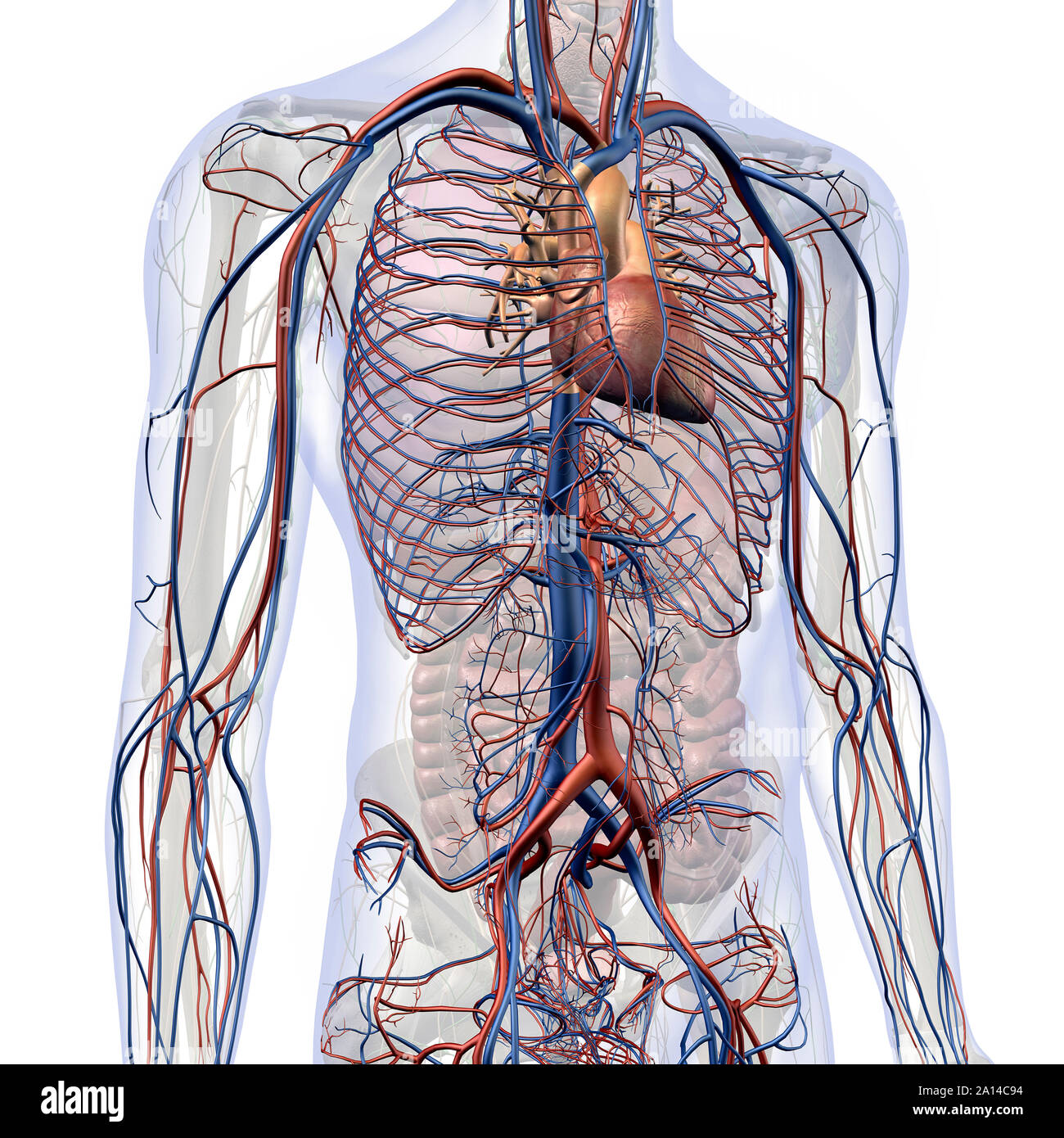
Male Internal Anatomy Of Heart And Circulatory System In Chest And Abdomen Stock Photo Alamy from c8.alamy.com The human thorax includes the thoracic cavity and the thoracic wall. The pectoralis major and the pectoralis minor, known collectively as your pecs. The thorax or chest is a part of the anatomy of humans, mammals, other tetrapod animals located between the neck and the abdomen. It provides protection to vital organs (eg, heart and major vessels, lungs, liver) and provides stability for movement. A good radiologist knows the anatomy because knowing where structures normally live and recognizing the location of an abnormality helps to make or narrow the differential diagnosis. A woman's chest — like the rest of her body — is covered with skin that has two layers. Chest a man's chest — like the rest of his body — is covered with skin that has two layers. Muscles of the chest and their functions you have two mighty muscles on both sides of your chest:
It is enclosed by the ribs, the vertebral column, and the sternum, or breastbone, and is separated from the abdominal cavity (the body's largest hollow space) by a muscular and membranous partition, the diaphragm.
Learn about each of these muscles, their locations, functional anatomy and exercises for them. Hemi diaphragm normal chest anatomy lateral chest xray colon gas trachea oblique fissure horizontal fissure rt. 31 anatomy of the female breast syllabus p. A woman's chest — like the rest of her body — is covered with skin that has two layers. It is enclosed by the ribs, the vertebral column, and the sternum, or breastbone, and is separated from the abdominal cavity (the body's largest hollow space) by a muscular and membranous partition, the diaphragm. 30 lines of the thoracic wall syllabus p. Knowledge of the anatomy of the whole cylinder (ribs, sternum, vertebra, diap … A good radiologist knows the anatomy because knowing where structures normally live and recognizing the location of an abnormality helps to make or narrow the differential diagnosis. Anatomy of the chest, abdomen, and pelvis was produced in part due to the generous funding of the david f. Here, we break down the anatomy of your chest muscles. The human thorax includes the thoracic cavity and the thoracic wall. The epidermis is the outermost layer that provides a protective, waterproof seal over the body. The circulatory system does most of its work.
Principal functions are the protection of internal viscera and an expandable cylinder facilitating variable gas flow into the lungs. Sternocleidomastoid muscle clavicle and ribs anatomy muscle anatomy chest sternocleidomastoid ribs anatomy chest muscles anatomy thorax rib muscles chest muscles chest anatomy illustration. Muscles of the chest and their functions you have two mighty muscles on both sides of your chest: A typical heart is approximately the size of your fist: The muscles of the chest develop from the somites found in the mesoderm.
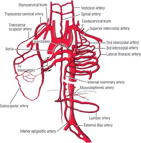
Applied Anatomy Of The Chest Wall And Mediastinum Basicmedical Key from basicmedicalkey.com The myotomes elongate and invade the mesoderm of the wall of the embryonic thoracic and abdominal cavities. This section of the website will explain large and minute details of arterial anatomy of chest The chest wall, like other regional anatomy, is a remarkable fusion of form and function. The epidermis is the outermost layer that provides a protective, waterproof seal over the body. Radiology basics of chest ct anatomy with annotated coronal images and scrollable axial images to help medical students and junior doctors learning anatomy. In insects, crustaceans, and the extinct trilobites, the thorax is one of the three main divisions of the creature's body, each of which is in turn composed of multiple segments. Anatomically, the heart is located in the anterior thoracic cavity; See human chest anatomy stock video clips.
Anatomy of the thorax, heart, abdomen and pelvis recommended text gray's anatomy for students.
12 cm (5 in) in length, 8 cm (3.5 in) wide, and 6 cm (2.5 in) in thickness. Chest muscles anatomy (1) pectoralis major muscle. Three dimensional view of the female reproductive system, full frontal view. Thoracic cavity, also called chest cavity, the second largest hollow space of the body. The pectoralis major and the pectoralis minor, known collectively as your pecs. Anatomically, the heart is located in the anterior thoracic cavity; A good radiologist knows the anatomy because knowing where structures normally live and recognizing the location of an abnormality helps to make or narrow the differential diagnosis. Learn about each of these muscles, their locations, functional anatomy and exercises for them. 2 skin of the anterior chest wall syllabus p. The muscles of the chest develop from the somites found in the mesoderm. The chest is made up primarily of two muscles: A woman's chest — like the rest of her body — is covered with skin that has two layers. Basic thoracic anatomy and physiology an understanding of thoracic imaging requires knowledge of the anatomy being imaged, as described in this chapter, as well as the imaging techniques applied to the thorax, covered in chapter 2.
Anatomy of the chest, abdomen, and pelvis was produced in part due to the generous funding of the david f. The chest wall is comprised of skin, fat, muscles, and the thoracic skeleton. The chest or thorax is the region between the neck and diaphragm that encloses organs, such as the heart, lungs, esophagus, trachea, and thoracic diaphragm. A line is drawn from anterior surface of the body of 6th thoracic vertebrae passing through the apex of the heart up to anterior lower most part of diaphragm. An overview of the anatomy visible in a transverse computed axial tomographical image of the thorax (and part of the abdomen) performed with intravenous cont.
Internal View Of The Chest Wall Anatomy Netter Illustration Used With Download Scientific Diagram from www.researchgate.net The chest wall is comprised of skin, fat, muscles, and the thoracic skeleton. 30 lines of the thoracic wall syllabus p. 31 anatomy of the female breast syllabus p. Basic thoracic anatomy and physiology an understanding of thoracic imaging requires knowledge of the anatomy being imaged, as described in this chapter, as well as the imaging techniques applied to the thorax, covered in chapter 2. The chest is the area of origin for many of the body's systems as it houses organs such as the heart, esophagus, trachea, lungs, and thoracic diaphragm. The chest anatomy includes the pectoralis major, pectoralis minor and the serratus anterior. Swensen fund for innovation in teaching. Knowledge of the anatomy of the whole cylinder (ribs, sternum, vertebra, diap …
4 innervation of the breast blood supply of the breast syllabus p.
Swensen fund for innovation in teaching. It is enclosed by the ribs, the vertebral column, and the sternum, or breastbone, and is separated from the abdominal cavity (the body's largest hollow space) by a muscular and membranous partition, the diaphragm. In insects, crustaceans, and the extinct trilobites, the thorax is one of the three main divisions of the creature's body, each of which is in turn composed of multiple segments. The anatomic illustrations are presented as… A typical heart is approximately the size of your fist: The chest wall is comprised of skin, fat, muscles, and the thoracic skeleton. These myotomes divide into the epimere and the hypomere. The human thorax includes the thoracic cavity and the thoracic wall. It provides protection to vital organs (eg, heart and major vessels, lungs, liver) and provides stability for movement. Basic thoracic anatomy and physiology an understanding of thoracic imaging requires knowledge of the anatomy being imaged, as described in this chapter, as well as the imaging techniques applied to the thorax, covered in chapter 2. Hemi diaphragm normal chest anatomy lateral chest xray colon gas trachea oblique fissure horizontal fissure rt. Three dimensional view of the female reproductive system, full frontal view. Learn about each of these muscles, their locations, functional anatomy and exercises for them.